Home » Without Label » Diagram Of Female Back - Female Anatomy Diagram Lower Abdomen | Stomach Pics ... / The back's muscles start at the top of the back (named the cervical vertebrae) and go to the tailbone (also named the coccyx).
Diagram Of Female Back - Female Anatomy Diagram Lower Abdomen | Stomach Pics ... / The back's muscles start at the top of the back (named the cervical vertebrae) and go to the tailbone (also named the coccyx).
Diagram Of Female Back - Female Anatomy Diagram Lower Abdomen | Stomach Pics ... / The back's muscles start at the top of the back (named the cervical vertebrae) and go to the tailbone (also named the coccyx).. The abdomen contains all the diagram of internal organs human body anatomy. This helps to balance and distribute the weight of the body on the legs. The red lines point individual bones and the names are writen in singular, the blue lines conect to group of bones and are in plural form. The vertebral column of the lower back includes the five lumbar vertebrae, the sacrum, and the coccyx. Female cardiovascular system, rear and front views, on black background.
Person outline body outline map outline body diagram body chart human body drawing body reference drawing free printable calendar templates body template. Female anatomy of cardiovascular system with skeleton. It is particularly interesting for physiotherapists. Posted on january 8, 2016 by admin. Female reproductive system 2021 | 4 minutes of easy learning mystery female body.
Back Muscles Diagram Female - Flexibility tips muscle ... from www.muscleblitz.com Female reproductive system 2021 | 4 minutes of easy learning mystery female body. The vertebral column of the lower back includes the five lumbar vertebrae, the sacrum, and the coccyx. Related posts of diagram of female back muscles muscle anatomy diagram. Related posts of diagram of a female lower back anatomy of pelvic bone. Posted on january 8, 2016 by admin. Anatomical diagrams of the spine and back. These two regions are responsible for most of the movement in the back, allowing. Anatomy of pelvic bone 12 photos of the anatomy of pelvic bone anatomical position of pelvic bone, anatomy and physiology of pelvic bone, anatomy of pelvic bone pdf, anatomy pelvic area bones, pelvic bone anatomy, human anatomy, anatomical position of pelvic bone, anatomy and physiology of pelvic bone, anatomy of.
The breadth of the back is created by the shoulders at the top and the pelvis at the bottom.
The kidneys lie retroperitoneally (behind the peritoneum) in the abdomen, either side of. Related posts of diagram of a female lower back anatomy of pelvic bone. The bones of the pelvis and lower back work together to support the body's weight, anchor the abdominal and hip muscles, and protect the delicate vital organs of the vertebral and abdominopelvic cavities. Anatomy of pelvic bone 12 photos of the anatomy of pelvic bone anatomical position of pelvic bone, anatomy and physiology of pelvic bone, anatomy of pelvic bone pdf, anatomy pelvic area bones, pelvic bone anatomy, human anatomy, anatomical position of pelvic bone, anatomy and physiology of pelvic bone, anatomy of. Urinary system of the lower torso. Every healthy human body has two kidneys, the left and the right. Other muscles are small and cover much less space. Female anatomy includes the external genitals, or the vulva, and the internal reproductive organs. Female reproductive system 2021 | 4 minutes of easy learning mystery female body. The human back, also called the dorsum, is the large posterior area of the human body, rising from the top of the buttocks to the back of the neck. The kidneys are some of the most important organs. These might range from a dull ache to a stabbing or shooting sensation. It is particularly interesting for physiotherapists.
Diagram of a human female skeleton, back view the red lines point individual bones and the names are writen in singular, the blue lines conect to group of bones and are in plural form. Download 2,913 body diagram female stock illustrations, vectors & clipart for free or amazingly low rates! Symptoms of low back pain. Most people have two kidneys, which are located near the back of the body, under the ribs, on each side of the spine.kidneys filter waste out of the bloodstream, which is passed out of the body as. The human back, also called the dorsum, is the large posterior area of the human body, rising from the top of the buttocks to the back of the neck.
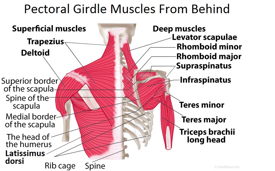
Pectoral Girdle Anatomy: Bones, Muscles, Function, Diagram ... from www.ehealthstar.com Other muscles are small and cover much less space. These two regions are responsible for most of the movement in the back, allowing. Bones of the pelvis and lower back. Lower back pain can be caused by a variety of problems in the lumbar spine. We are pleased to provide you with the picture named anatomy of back muscles diagram. The back consists of the spine, spinal cord, muscles, ligaments, and nerves. Some of these muscles are quite large and cover broad areas. The back's muscles start at the top of the back (named the cervical vertebrae) and go to the tailbone (also named the coccyx).
This diagram depicts female human anatomy 744×1116 with parts and labels.
The pelvis at the bottom of the back and the shoulders at the top of the back give the back its breadth, and it narrows in between these two regions. The abdomen contains all the diagram of internal organs human body anatomy. See back muscle anatomy stock video clips. See human back anatomy stock video clips. Anatomy of pelvic bone 12 photos of the anatomy of pelvic bone anatomical position of pelvic bone, anatomy and physiology of pelvic bone, anatomy of pelvic bone pdf, anatomy pelvic area bones, pelvic bone anatomy, human anatomy, anatomical position of pelvic bone, anatomy and physiology of pelvic bone, anatomy of. Diagram of a human female skeleton, back view the red lines point individual bones and the names are writen in singular, the blue lines conect to group of bones and are in plural form. It is particularly interesting for physiotherapists. It is the surface of the body opposite from the chest and the abdomen.the vertebral column runs the length of the back and creates a central area of recession. We hope this picture anatomy of back muscles diagram can help you study and research. The human back extends from the buttocks to the posterior portion of the neck and shoulders.it is opposite from the chest, and the vertebral column runs down the back. The kidneys are some of the most important organs. That means it's really sensitive, and for many women, stimulating it is the best way to orgasm. Cervical spine diagram stress in the spine is greatest in the cervical (neck) and lumbar (lower back) areas.
Diagram of a human female skeleton, back view the red lines point individual bones and the names are writen in singular, the blue lines conect to group of bones and are in plural form. This diagram depicts female human anatomy 744×1116 with parts and labels. The first is to produce egg cells, and the second is to protect and nourish the offspring until birth. Diagram of a human female skeleton, back view. Urinary system of the lower torso.
Urinary System Female | Anatomy organs, Anatomy flashcards ... from i.pinimg.com The human back, also called the dorsum, is the large posterior area of the human body, rising from the top of the buttocks to the back of the neck. Human musculature bodybuilding infographic muscular system vector human anatomy back muscle anatomy bicep male muscular anatomy human body anatomy female female anatomy muscle hamstrings muscle. Certain back muscles extend to other areas, like the shoulders, upper arms, and thighs. It is the surface of the body opposite from the chest and the abdomen.the vertebral column runs the length of the back and creates a central area of recession. These structures work together to support the body, enable a range of movements, and send messages from the brain to. Person outline body outline map outline body diagram body chart human body drawing body reference drawing free printable calendar templates body template. An illustration showing a theory of vision published in treatise on man by rene descartes. This helps to balance and distribute the weight of the body on the legs.
See lower back pain symptoms, diagnosis, and treatment.
We hope this picture anatomy of back muscles diagram can help you study and research. We are pleased to provide you with the picture named anatomy of back muscles diagram. Diagram of a human female skeleton, back view the red lines point individual bones and the names are writen in singular, the blue lines conect to group of bones and are in plural form. Female reproductive system 2021 | 4 minutes of easy learning mystery female body. The abdomen contains all the diagram of internal organs human body anatomy. The human back, also called the dorsum, is the large posterior area of the human body, rising from the top of the buttocks to the back of the neck. Cervical spine diagram stress in the spine is greatest in the cervical (neck) and lumbar (lower back) areas. Certain back muscles extend to other areas, like the shoulders, upper arms, and thighs. The human back extends from the buttocks to the posterior portion of the neck and shoulders.it is opposite from the chest, and the vertebral column runs down the back. Anatomy of pelvic bone 12 photos of the anatomy of pelvic bone anatomical position of pelvic bone, anatomy and physiology of pelvic bone, anatomy of pelvic bone pdf, anatomy pelvic area bones, pelvic bone anatomy, human anatomy, anatomical position of pelvic bone, anatomy and physiology of pelvic bone, anatomy of. These two regions are responsible for most of the movement in the back, allowing. Each kidney is about four or five inches long. The pelvis at the bottom of the back and the shoulders at the top of the back give the back its breadth, and it narrows in between these two regions.